Everything we have studied so far ends up here. This is the interpretation level of the four levels of description, and also the place where the ladder folds back on itself: expectation and attention change what the auditory cortex does to a signal, so the highest level reaches down and alters the lowest. The cochlea and psychoacoustics, embodiment, and physiological reactions all feed a brain that turns vibrating air into pitch, rhythm, timbre, emotion, and memory. The signal is reshaped at every step as it travels from the ear to the cortex and the limbic system. Harmony and melody described pitch and tonal structure in musical terms; neuroscience often takes those same dimensions into the laboratory and manipulates them. This week looks at which brain regions are doing the work, how they cooperate, and how researchers measure their activity while we listen and perform.
The human brain
Some 86 billion neurons, plus an even larger number of supporting glial cells, make the brain the most intricate organ we have. It is where all sensory input is gathered and where perception, cognition, emotion, and movement are coordinated. The brain is also remarkably plastic. It reorganises itself in response to musical training, sensory experience, and even injury. That is why we can pick up new musical skills, adapt after hearing loss, and grow particular musical tastes. By weaving together signals from the outside world and from our own internal states, it lets us interpret sound and music and find meaning in them. Its main anatomical regions include:
Frontal lobe: At the front of the brain, this region handles executive functions—decision-making, planning, problem-solving, voluntary movement—and shapes aspects of personality and social behaviour.
Parietal lobe: Sitting behind the frontal lobe, it processes bodily sensation: touch, temperature, pain, and spatial orientation.
Temporal lobe: Tucked beneath the frontal and parietal lobes, it carries much of our auditory processing, language comprehension, and memory formation.
Occipital lobe: At the back of the brain, this region is devoted mainly to vision.
Cerebellum: Below the occipital lobe, it coordinates voluntary movement, balance, posture, and motor learning.
Cingulate gyrus: A part of the limbic system, it contributes to emotion, learning, and memory, and ties behavioural outcomes to motivation.
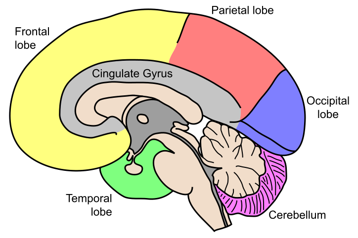
Illustration of the brain’s lobes (Wikimedia Commons).
Neurons
Neurons are the basic units of the brain and nervous system. These specialised cells carry information around the body using both electrical and chemical signals, from simple reflexes to thoughts and emotions. A neuron has a few main parts: the cell body, or soma which holds the nucleus and the cell’s machinery; dendrites, the branch-like extensions that receive signals from other neurons; and the axon, a long projection that carries electrical impulses away from the cell body towards other neurons, muscles, or glands. Neurons communicate at the synapse, a small gap where chemical messengers called neurotransmitters pass the signal from the axon terminal of one neuron to the dendrite of another.
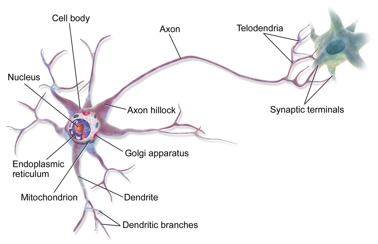
Illustration of a multipolar neuron (Wikimedia Commons).
The exchange starts when dendrites pick up signals from neighbouring neurons. The cell body adds these inputs together, and if the total is strong enough it fires an action potential, an electrical impulse that travels down the axon. When that impulse reaches the synapse, neurotransmitters are released and the signal moves on to the next neuron. Repeated across billions of cells, this is how the brain processes sensory information, controls movement, and generates thoughts, emotions, and memories.
Dig deeper: artificial neural networks
The brain has also served as a design metaphor. Artificial neural networks (ANNs), the machinery behind most of what is now called artificial intelligence, take their name and their basic picture from the cells described above: many simple units, each summing its inputs and passing on a result, wired together in layers whose connection strengths change with use.
The analogy is worth holding loosely. A network’s “learning” is the adjustment of numerical weights to reduce an error, which is a thin cartoon of synaptic plasticity, and its units have none of the chemistry, timing, or anatomy of a neuron. The traffic between the two fields has nonetheless run both ways. Neuroscience lent machine learning a metaphor, and trained networks are now used in return as explicit hypotheses about what the auditory system might be computing, to be tested against recordings from real brains. Machine listening takes the topic up on the technical side.
The listening brain
Hearing begins in the ear, but listening is done by the brain. The regions and pathways below are the ones that turn the signal from the cochlea into a heard sound, and then into a melody, a beat, a memory, and a feeling.
Key brain regions for listening
A handful of regions do much of the work when we process sound and music:
- The auditory cortex, in the temporal lobe, decodes basic sound features such as pitch, loudness, and timbre, and is central to recognising and interpreting musical elements.
- The prefrontal cortex handles higher-order work: directing attention, spotting patterns, and predicting where the music is going.
- The motor cortex lights up even when we only listen, reflecting how the brain responds to rhythm and beat and prepares for movement Zatorre et al., 2007.
- The limbic system, which includes the amygdala and hippocampus, drives emotional responses to music and ties pieces to memories.
- The nucleus accumbens, part of the reward system, is linked to the pleasure and motivation that music can stir up Salimpoor et al., 2011.
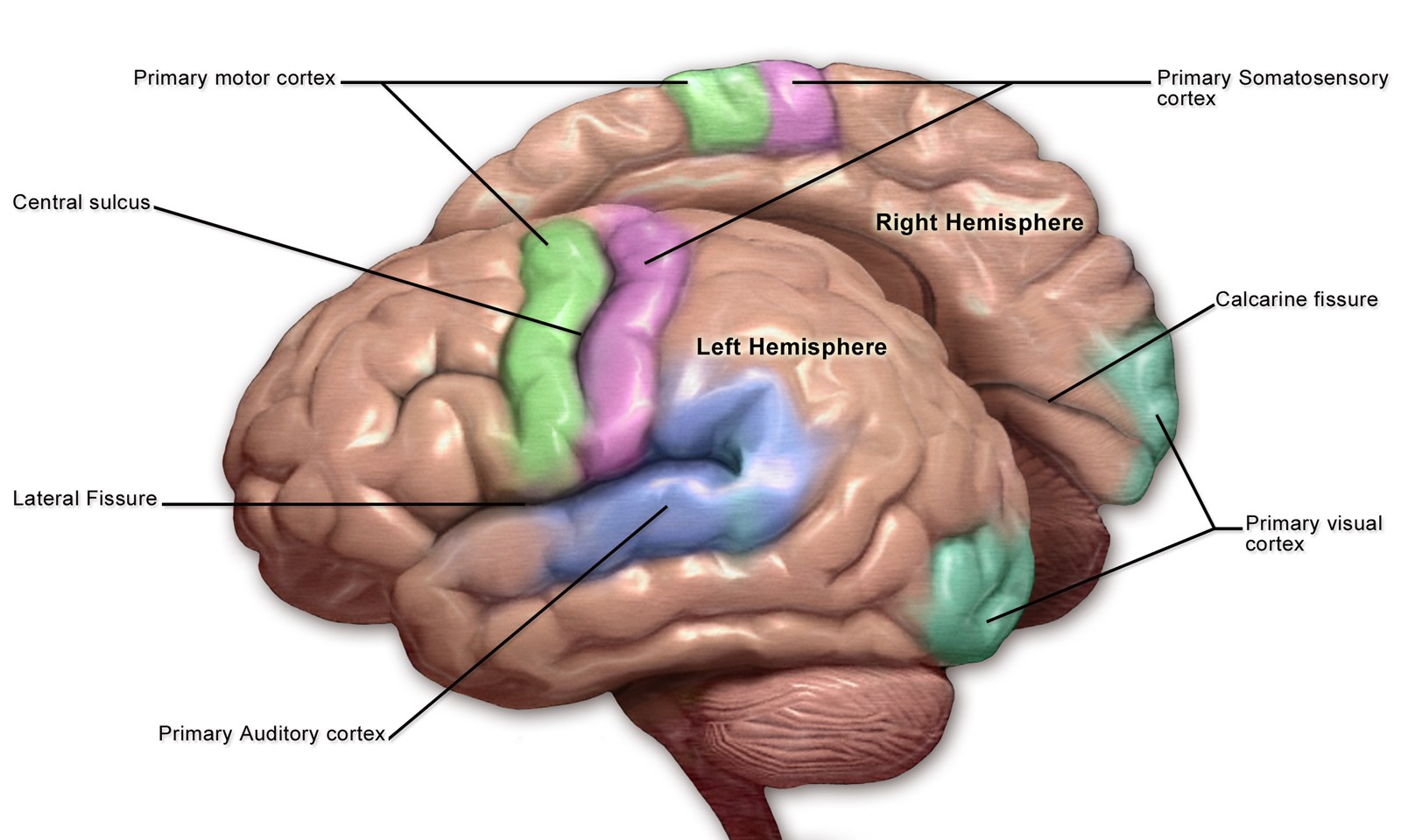
Motor and Sensory Regions of the Cerebral Cortex (Illustration: Blausen Medical).
Auditory pathways
Psychoacoustics followed sound as far as the cochlea, where hair cells turn movement into nerve impulses along a tonotopic map. This chapter picks the signal up from there. The auditory nerve carries it to the brainstem and its first synapse in the cochlear nuclei, and from there it climbs through several relay stations in the brainstem, midbrain, and thalamus to the cortex.
Dig deeper: the relay stations of the auditory pathway
- The superior olivary complex, in the brainstem, compares the timing and intensity of sound at the two ears, which is the basis for locating sounds in space.
- The inferior colliculus, in the midbrain, gathers auditory information and supports reflexive responses such as turning the head towards a noise.
- The medial geniculate nucleus of the thalamus works as a relay hub, routing auditory signals on to the right parts of the cerebral cortex.
The signal finally arrives at the primary auditory cortex in the temporal lobe, where basic features such as pitch, loudness, and timbre are decoded. Secondary auditory areas and other cortical regions take it from there, letting us recognise complex sounds, speech, and music. Patel (2007) is the standard treatment of how far the brain’s machinery for music and for language overlaps.
Because the system processes sound in both a hierarchical and a parallel fashion, the brain can analyse several aspects of a sound at once. That is what lets us detect, locate, and make sense of everything from a single voice to the layered patterns of music.
Neural processing of sound
Once auditory information reaches the brain, specialised circuits set about decoding it. In the auditory cortex, different populations of neurons are tuned to particular frequencies, which is what lets us tell musical notes and speech sounds apart:
- Pitch: Neurons in the primary auditory cortex are arranged tonotopically—laid out according to the frequency they respond to—so the brain can tell high pitches from low.
- Loudness: Intensity is coded in how fast auditory neurons fire; louder sounds drive stronger responses.
- Timbre: The colour or quality of a sound comes from combining many frequencies and harmonics, which is how we distinguish one instrument or voice from another.
- Rhythm and timing: Temporal patterns in music and speech are tracked by networks spanning the auditory cortex and motor-related areas, supporting our knack for hearing and moving with a beat.
Dig deeper: lateralisation
Lateralisation describes how some auditory functions lean more on one hemisphere than the other. The left hemisphere tends to specialise in rapid temporal changes, like those in speech and rhythm, which makes it important for language comprehension and rhythmic analysis. The right hemisphere is generally more sensitive to spectral, frequency-based information, and so matters more for melody, pitch, and the emotional shading of music. Splitting the work this way lets the brain analyse complex sounds like music by handling different features in parallel.
Once the features are extracted, they are folded together with input from other regions: the prefrontal cortex for attention and expectation, the limbic system for emotional response, and the motor cortex for rhythm and motion. This is the stage where we recognise a familiar tune, sense where the music is headed, and link a sound to a memory or a feeling.
Music cognition
Music cognition is about the mental work of understanding, interpreting, and responding to music. Listening pushes the brain well past basic sound analysis: we recognise melodies, anticipate harmonic moves, feel rhythm, and call up musical memories. Several regions share this load:
- The prefrontal cortex, which recognises patterns, sets up musical expectations, and ties the music together with attention and working memory.
- The motor cortex, which comes into play for rhythm perception, beat tracking, synchronisation, and the planning of music-related movement.
- The limbic system, with the amygdala and hippocampus, which connects music to emotions and autobiographical memories and gives a piece its personal weight.
- The reward system, especially the nucleus accumbens, which fires during pleasurable or meaningful moments and feeds motivation and enjoyment.
Linked up like this, these networks let us follow musical structure, anticipate what comes next, feel something, and tie music to our own past. How exactly these systems cooperate is still an active research question, and it is what underlies our unusual capacity to find meaning in music.
Memory, attention, expectation, learning, and development
Perception is only the way in. Listening to music keeps memory, attention, expectation, and learning busy the whole time. These processes are spread across the pathways described above—especially the prefrontal and parietal cortices, the auditory areas, and the limbic structures—and they shift with age, training, and culture (see also listening on enculturation and expertise).
Memory
A useful simplification splits memory three ways: sensory memory (the very brief persistence of a sound), working memory (actively holding and reshaping a short musical pattern such as a melodic fragment, a chord change, or a rhythmic grouping), and long-term memory (knowledge and episodes built up over time). Long-term memory itself is often divided into explicit memory (conscious recall: that tune, those lyrics) and implicit memory (skills and expectations you never set out to memorise, like sensing a “wrong” note in a familiar style).
In musical terms, the hippocampus and the cortex around it support the recognition and recall of familiar material and its autobiographical associations, which is why a few seconds of sound can work like a time machine. Meanwhile the statistical regularities of the music you grew up with quietly shape your expectations about melody and harmony, without your ever naming the rules. That kind of learning does not even need attention: intracranial recordings from patients who were reading while random tone sequences played in the background show that the brain kept encoding the statistical structure of the sequences anyway, in a network reaching well beyond the auditory cortex Fuhrer et al., 2025. Earworms, those involuntary loops of musical imagery, are a reminder that musical memories can be triggered and run on with a will of their own.
Attention
Attention decides which streams and features matter right now. Selective attention lets you lock onto one instrument or voice in a mixture (the cocktail-party problem from psychoacoustics); divided attention is what you need when several parts demand watching at once. The cues can be endogenous (you choose what to listen for) or exogenous (a sudden change in timbre grabs you). Overt attention includes where you point your eyes—relevant when reading a score or watching performers (vision)—while covert attention shifts the “listening spotlight” with the eyes held still.
Frontal and parietal networks bias what the auditory cortex does, which is why so many EEG and fMRI studies manipulate attention on purpose. The same acoustic stimulus can produce different brain responses depending on the task and where the listener’s focus sits.
Expectation
Listeners do not just hear what arrives; they hear it against predictions about what is likely to come next. Exposure builds an internal model of pitch, timing, and style, and surprise—a violation of those predictions—is a major source of tension, humour, and emotion in music (which connects naturally to harmony and melody). Psychologically this shades into statistical learning. You soak up the probabilities of how things continue without anyone teaching them.
Dig deeper: event-related potentials and the mismatch negativity
In cognitive neuroscience, event-related potentials (ERPs) such as the mismatch negativity (MMN) have been used to track the automatic detection of rule-breaking or rare sounds—even when listeners are not paying attention to the stream—while other components reflect more conscious expectation and updating.
Expectation thus bridges low-level auditory processing and the richer descriptions of music theory. Rhythm gives expectation a clock. EEG work at the University of Oslo shows that listeners perceive a sound better when it arrives at an expected moment, and that strict periodicity and learnable irregular patterns each contribute to that expectation Solli et al., 2025.
Learning and development
Neuroplasticity means experience rewires synapses. Musical training is linked to differences in auditory, motor, and multimodal regions, and to how tightly the motor system couples to heard rhythms. Development adds the dimension of time. Infants and children are tuned to the musical features of their surroundings early on, and some capacities—certain aspects of pitch learning, for instance—show sensitive periods, windows in which experience leaves an outsized lasting mark. That does not mean adults learn nothing.
Across a lifetime, enculturation—the statistical structure of the music around you—sets what counts as “normal” or surprising, and individual differences in training, personality, and neurodiversity colour how these systems respond. Taken together, memory, attention, expectation, and learning explain why the same score or recording can be processed so differently by two brains, and why longitudinal and cross-cultural studies matter alongside laboratory experiments.
Music, emotion, and aesthetic experience
Sound and music draw on reward, memory, and motivation circuits, not just the “cold” analysis of pitch and time. Neuroscientists often separate three things:
- Core affect — quick changes in valence and arousal tied to acoustic surprise or familiarity.
- Episodic memories — music as a time machine for autobiographical scenes.
- Aesthetic judgement — reflective appraisal (preference, beauty, interestingness) that can diverge from immediate pleasure.
The phenomena that matter musically include chills, the nostalgia a snippet of melody can set off, and moving performances that lean as much on timing and timbre as on pitch structure. All of this connects auditory cortex and brainstem pathways to limbic and prefrontal regions Koelsch, 2014, so imaging and EEG studies have to read emotion-related findings carefully. Many designs mix expressive cues (the performance gestures you met in the embodiment chapter) with purely auditory signals.
For musicology, keep three distinctions in play:
- Psychoacoustic salience versus cultural framing: the same acoustic change can mean different things across repertoires.
- Laboratory emotion scales versus historically situated aesthetics: what critics or philosophers actually describe.
- Group averages versus individual listeners: a distinction that matters especially for clinical-adjacent claims.
These themes tie perception (psychoacoustics), the moving and listening body (previous chapters), and the critical reading of empirical papers (Reading empirical research in the introduction) back together.
Mirror neurons
Mirror neurons are a special class of cell that fire both when you perform an action and when you watch someone else perform the same one. First found in the premotor cortex of monkeys, they have since been identified in humans and are thought to matter for understanding actions, imitation, empathy, and social cognition Rizzolatti & Craighero, 2004. In music, mirror-neuron systems are believed to help us take in and internalise musical gestures, rhythms, and emotions. Watch a musician play or a dancer move and your mirror neurons may fire as though you were doing it yourself. That internal echo supports learning by imitation, but also the embodied, emotional side of music. That is part of why we can “feel” the beat or read a performer’s expression as our own. It also fits the picture from the psychoacoustics chapter: listeners hear sounds as actions on objects, drawing on internalised action–sound couplings, and the motor system appears to take part in that hearing.
That account is popular, and it is also contested. Three caveats are worth carrying with you:
- The direct evidence in humans is thin. Mirror neurons were found by recording from single cells in macaques. That is rarely possible in humans, so most human claims rest on indirect measures—fMRI or EEG showing that motor areas are active during observation—which cannot show that the same neurons do both jobs.
- Activation is not understanding. Critics, Gregory Hickok most prominently Hickok, 2009, argue that motor activity during observation may be a consequence of understanding an action rather than its cause; people with damage to motor regions can still recognise actions they can no longer perform.
- The leap to empathy and music is large. Going from “this neuron fires when the monkey grasps and when it sees grasping” to “this is why we feel a performer’s expression” crosses several inferential steps that the data do not license on their own.
None of this means the mirror-neuron literature is worthless. The coupling between listening and motor systems is robust and shows up throughout this course. It means the mechanism is still argued over, and that “mirror neurons explain it” is a hypothesis rather than a finding.
Individual differences
People vary widely in how they hear and process music, shaped by genetics, development, experience, and culture. Some are naturally more sensitive to pitch, rhythm, or timbre; others have unusual abilities such as absolute pitch or unusually strong emotional reactions. How strongly people are moved by music is itself partly heritable. In a study of about 2,600 Norwegian twins, musical sensibility was linked to openness (especially aesthetic sensitivity), agreeableness, and negative emotionality, and those links were driven mainly by shared genes Hansen et al., 2025. Training counts for a lot here, since it can boost neural connectivity and plasticity across auditory, motor, and cognitive regions. Musicians often show sharper auditory discrimination, better memory for musical patterns, and tighter integration of sensory and motor information. Their training can strengthen the links between the hemispheres in ways that support demanding skills like sight-reading or improvising.
Age matters too. As we get older, changes in the auditory system and the brain can blunt sensitivity to some frequencies and make it harder to pick out speech or fine musical detail. But neuroplasticity allows for compensation, and staying musically active across life helps keep auditory and cognitive functions in good shape.
Neurodiversity adds further variation. People with autism spectrum disorder (ASD), for instance, may process music differently, sometimes with enhanced pitch perception or distinctive emotional responses. At the other end, amusia (musical tone deafness) can limit the perception of pitch or rhythm even when hearing and intelligence are otherwise unaffected.
Cultural background and exposure leave their mark as well. Early experiences with the music, language, and rhythm of a particular culture shape neural pathways and tastes, and colour how we hear unfamiliar styles later on.
What runs through all of this is the brain’s adaptability. Through training, experience, and adjustment to sensory challenges, it keeps reorganising itself, which is what lets us learn new musical skills, adapt after hearing loss, and develop our own preferences and abilities.
Capturing brain activity
To study how the brain handles sound and music, researchers need ways to measure its activity. A handful of non-invasive techniques are in common use, in both research and the clinic, for watching neural responses during listening and performance. Each one tells us something different about the timing, the location, and the nature of what the brain is doing.
EEG (electroencephalography)
Electroencephalography (EEG) records the brain’s electrical activity through electrodes placed on the scalp. These pick up the tiny voltage fluctuations produced when large groups of neurons, mostly in the cerebral cortex, fire together. EEG is especially good at catching fast, millisecond-scale changes, which makes it well suited to studying the timing and dynamics of brain responses.
In sound and music research, EEG is used to follow how the brain handles rhythm, pitch, melody, and harmony. Present a musical stimulus and you can observe event-related potentials (ERPs), which are distinctive patterns in the EEG signal that are time-locked to a specific sensory, cognitive, or motor event. This opens a window onto auditory attention, expectation, and memory as they happen.
EEG is also a clinical workhorse, used to diagnose conditions such as epilepsy, sleep disorders, and brain injuries. In music neuroscience it has helped show how musical training reshapes brain function, how the brain tells musical genres apart, and how emotional responses to music arise.
Its strengths are excellent temporal resolution on the millisecond scale, non-invasiveness, relatively low cost, and portability, all of which make it usable with many kinds of participant and setting. The trade-offs are limited spatial resolution—it is hard to pin down exactly where a signal comes from—and a bias towards neurons near the scalp. EEG recordings are also vulnerable to artefacts from muscle movement and eye blinks.
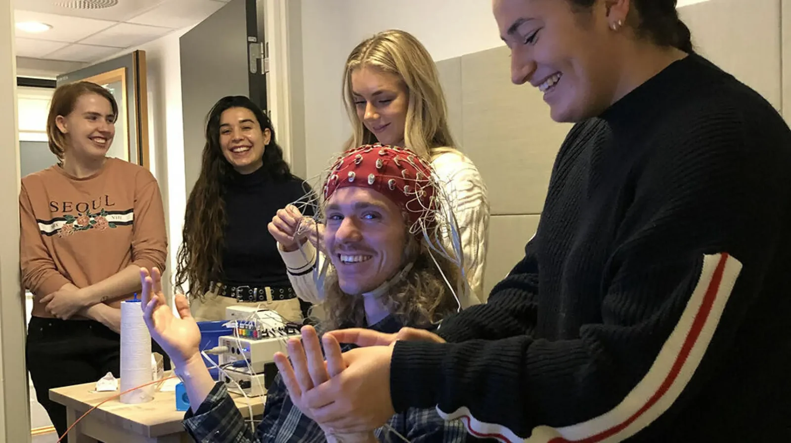
Research assistants and students are practising EEG measurement at RITMO (Photo: UiO).
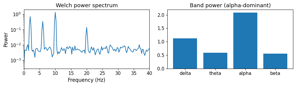
EEG is usually summarised by the power in standard frequency bands, named delta, theta, alpha, and beta, running from the slowest waves to the fastest. Here we synthesise a signal dominated by alpha waves, estimate its power spectrum with Welch’s method (a standard averaging technique), and read off the band powers, which is the same first step used in real analyses.
Source
import numpy as np
from scipy.signal import welch
import matplotlib.pyplot as plt
plt.rcParams.update({
    "font.size": 10,
    "axes.titlesize": 11,
    "axes.labelsize": 10,
    "xtick.labelsize": 9,
    "ytick.labelsize": 9,
    "legend.fontsize": 9,
    "figure.figsize": (10, 4),
    "figure.dpi": 100,
    "savefig.dpi": 150,
    "axes.spines.top": False,
    "axes.spines.right": False,
})

rng = np.random.default_rng(0)
fs = 256
t = np.arange(0, 20.0, 1 / fs)
sig = (1.0 * np.sin(2 * np.pi * 10 * t)    # alpha (strong)
       + 0.5 * np.sin(2 * np.pi * 6 * t)   # theta
       + 0.3 * np.sin(2 * np.pi * 20 * t)  # beta
       + 0.7 * np.sin(2 * np.pi * 2 * t)   # delta
       + 0.8 * rng.standard_normal(len(t)))

f, P = welch(sig, fs=fs, nperseg=1024)
bands = {"delta": (0.5, 4), "theta": (4, 8), "alpha": (8, 13), "beta": (13, 30)}
powers = {name: P[(f >= lo) & (f < hi)].sum() for name, (lo, hi) in bands.items()}

fig, ax = plt.subplots(1, 2, figsize=(10, 3))
ax[0].semilogy(f, P)
ax[0].set_xlim(0, 40)
ax[0].set_xlabel("Frequency (Hz)")
ax[0].set_ylabel("Power")
ax[0].set_title("Welch power spectrum")
ax[1].bar(list(powers.keys()), list(powers.values()))
ax[1].set_title("Band power (alpha-dominant)")
plt.tight_layout()
plt.show()
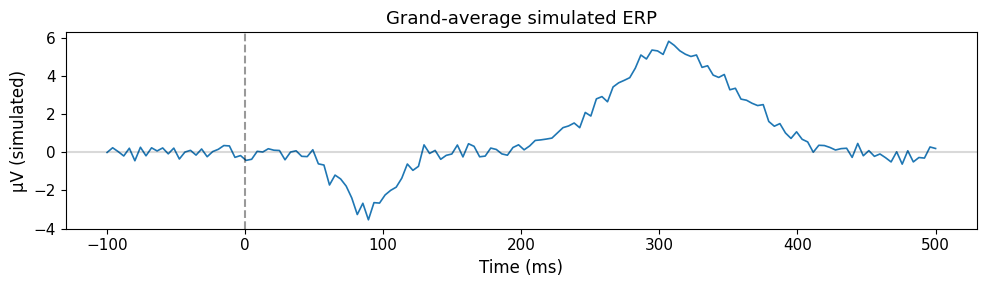
EEG is also used to study the response to single events, such as a chord or a note. Each response is tiny next to the ongoing activity, so researchers present the same event many times and average the recordings: the ongoing activity cancels out, and the time-locked response, an event-related potential (ERP), emerges. The simulation below shows this happening with synthetic data.
Source
import numpy as np
import matplotlib.pyplot as plt
plt.rcParams.update({
    "font.size": 10,
    "axes.titlesize": 11,
    "axes.labelsize": 10,
    "xtick.labelsize": 9,
    "ytick.labelsize": 9,
    "legend.fontsize": 9,
    "figure.figsize": (10, 4),
    "figure.dpi": 100,
    "savefig.dpi": 150,
    "axes.spines.top": False,
    "axes.spines.right": False,
})

sr_eeg = 250
t_ms = np.linspace(-100, 500, int(600 * sr_eeg / 1000), endpoint=True)
n_trials = 100
rng = np.random.default_rng(1)
avg = np.zeros_like(t_ms)
for _ in range(n_trials):
    trial = 2.5 * rng.standard_normal(len(t_ms))
    trial -= 3.0 * np.exp(-((t_ms - 90) ** 2) / (2 * 18**2))
    trial += 5.5 * np.exp(-((t_ms - 310) ** 2) / (2 * 45**2))
    avg += trial
avg /= n_trials

fig, ax = plt.subplots(figsize=(10, 3))
ax.plot(t_ms, avg, lw=1.2)
ax.axvline(0, color="k", linestyle="--", alpha=0.4)
ax.axhline(0, color="k", linestyle="-", alpha=0.15)
ax.set_xlabel("Time (ms)")
ax.set_ylabel("µV (simulated)")
ax.set_title("Grand-average simulated ERP")
plt.tight_layout()
plt.show()
MEG (magnetoencephalography)
Magnetoencephalography (MEG) measures the faint magnetic fields thrown off by synchronised electrical activity in groups of neurons, again mostly in the cerebral cortex. Where EEG reads voltage changes at the scalp, MEG reads the magnetic signals from neural currents directly, giving a clean view of brain function with high temporal precision.
This makes MEG especially valuable for studying the timing and location of the processes behind sound and music perception. Its millisecond-level resolution lets researchers track the fast neural dynamics of auditory processing, rhythm perception, and musical imagery. It can also map functional connectivity—how regions talk to each other during a musical task—by looking at how neural activity synchronises across the cortex.
In music neuroscience, MEG has been used to study how the brain distinguishes musical elements, how musicians anticipate and lock onto rhythm, and how emotional responses unfold over time. Because it localises sources more accurately than EEG, it is a strong choice for exploring the spatial layout of auditory and cognitive functions.
Its strengths are excellent temporal resolution on the millisecond scale, good spatial resolution, and direct, non-invasive measurement of neural activity, and it is particularly good at mapping connectivity between regions. The catches are real, though: MEG is expensive, needs a magnetically shielded room to keep out interference, is less portable and more movement-sensitive than EEG or fNIRS, and is harder to access because of the specialised equipment it requires.
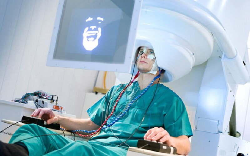
MEG instrument used for recording magnetic fields generated by brain activity (Photo: Very Big Brain).
fMRI (functional magnetic resonance imaging)
Functional Magnetic Resonance Imaging (fMRI) tracks brain activity indirectly, by detecting changes in blood flow, specifically the Blood Oxygen Level Dependent (BOLD) signal. When a region becomes more active it uses more oxygen, which shifts the local blood oxygenation in ways fMRI can pick up. From this it builds detailed maps of activity across the whole brain.
In music neuroscience, fMRI is the go-to for asking which areas are engaged during listening, performance, and imagining music. It has shown how networks for auditory perception, emotion, memory, and motor planning respond to music. Listening, for example, engages not only the auditory cortex but also limbic regions (emotion), the prefrontal cortex (attention and expectation), and motor areas (rhythm and movement).
The strength of fMRI is its spatial resolution. It localises activity precisely. The cost is temporal resolution, which is lower than EEG or MEG because the blood-flow (hemodynamic) response plays out over several seconds. The scanner is also loud and demands that participants hold very still, which limits the kinds of musical task you can run.
In short, fMRI offers high spatial resolution on the millimetre scale, whole-brain coverage, and non-invasive measurement, and it is widely available and well suited to mapping complex networks. Against that, its temporal resolution is on the order of seconds, it is sensitive to head motion, the environment is noisy and confined, and it is expensive. It is also off-limits for some people, including those with metal implants or claustrophobia.
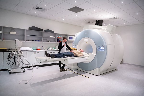
Example of an fMRI scan (Image: Wikimedia Commons).
fNIRS (functional near-infrared spectroscopy)
Functional Near-Infrared Spectroscopy (fNIRS) shines near-infrared light through the scalp to monitor changes in blood oxygenation and blood volume in the cortex, which are indirect signs of neural activity. Light sources and detectors on the head measure how much light is absorbed by oxygenated and deoxygenated haemoglobin. When a region becomes more active and uses more oxygen, the optical properties of the tissue change in a detectable way.
fNIRS comes into its own where other methods are awkward. Because it is silent and tolerates movement, it suits work with infants, children, and musicians playing their instruments, as well as experiments in more naturalistic, real-world settings. It has been used to study how children process music, how musicians’ brains respond during live performance, and how social interaction shapes neural activity in group music-making.
Its strengths are portability, relatively low cost, silent operation, and movement tolerance, which open it up to a wide range of participants and settings, including developmental and ecological studies. Its main limits are that it reaches only cortical (surface) regions, has lower spatial resolution than fMRI, and cannot see deep brain structures.
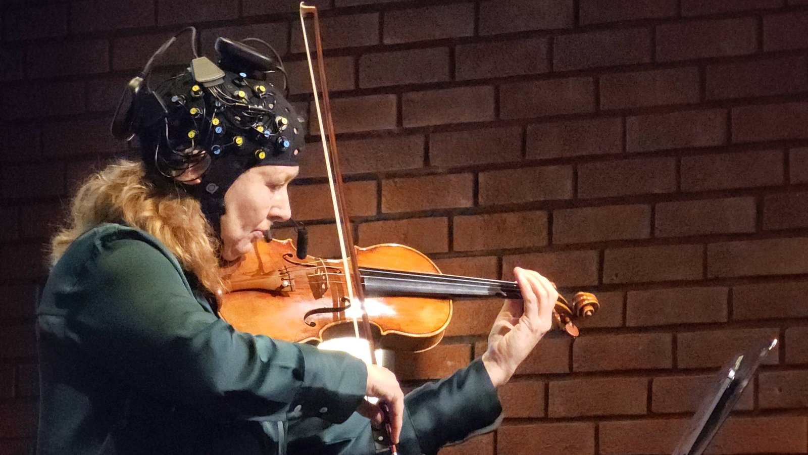
Victoria Johnson performing with an fNIRS system during the event MusicLab Brain: Inside the mind of a violinist in 2024 (Photo: UiO).
Comparison of brain activity measurement methods
Taken together, EEG, fNIRS, MEG, and fMRI cover the whole range, from the millisecond timing of neural events to the precise location of the regions behind perception, cognition, and emotion. Each makes different compromises, so the choice of method depends on the question.
| Method | Typical sampling | Strengths | Limitations |
|---|---|---|---|
| EEG | 250–1,000 Hz (up to several kHz in research systems) | Excellent temporal resolution (milliseconds); non-invasive; affordable; portable; suitable for diverse participants and settings | Limited spatial resolution; signals mainly from surface neurons; susceptible to artefacts (e.g., muscle movement, eye blinks) |
| fNIRS | around 10 Hz | Portable; affordable; silent; tolerant of movement; suitable for developmental and ecological studies | Limited to cortical (surface) regions; lower spatial resolution than fMRI; cannot access deep brain structures |
| MEG | around 1,000 Hz | Excellent temporal resolution; good spatial resolution; direct measurement of neural activity; non-invasive; suitable for mapping connectivity | High cost; requires magnetically shielded room; less portable; sensitive to movement; limited accessibility |
| fMRI | one whole-brain image every 1–2 s (about 0.5–1 Hz) | High spatial resolution (millimetres); whole-brain coverage; non-invasive; widely available; effective for mapping complex networks | High cost; lower temporal resolution (seconds); sensitive to head movement; noisy/confined environment; not suitable for all populations |
The sampling column follows the same logic as the physiology table in the previous chapter: the rate must comfortably exceed twice the fastest change of interest. Neural oscillations reach into the gamma band, roughly 30–100 Hz, so EEG and MEG sample at hundreds to thousands of hertz. fMRI does not sample electrical activity at all but a slow blood-oxygenation response that unfolds over several seconds, so one image every second or two is enough, and no faster scanner would change that limit.
Clinical and creative applications
The methods above were developed to observe the brain. Two fields use them to act on it or with it: brain-computer interfaces, which turn brain activity into control signals, and music therapy, which uses music to change what the brain does.
Brain-computer interfaces
Brain-computer interfaces (BCIs) let the brain communicate with external devices directly, sidestepping the usual route through muscles or speech. For sound and music, this opens up a range of uses that tap neural activity to control or shape what we hear.
Brain-computer interfaces for music: BCIs can let people create or control music with their brain signals. EEG-based systems, for instance, allow users to compose melodies, trigger musical events, or adjust sound parameters through imagined movement or focused attention. These are new routes to musical expression, especially for people with physical disabilities.
Hearing aids: Modern hearing aids already adapt their signal processing to the surrounding environment. Researchers are now asking whether BCIs can go further by reading the user’s auditory attention, so the device can foreground the sound the listener actually wants, such as a particular voice in a noisy room.
Cochlear implants: Cochlear implants restore hearing by stimulating the auditory nerve directly in people with severe hearing loss. Work in neuroscience and brain-computer interfacing is leading towards smarter implants that respond to the user’s own neural activity, with the promise of better sound quality and clearer speech.
Across these examples the same idea recurs. Reading and working with brain activity can sharpen auditory perception, communication, and creative expression. As the technology matures, BCIs and neuroprosthetic devices look set to matter more and more, in clinical and artistic work alike.
Music therapy
The clinical thread running through hearing aids and cochlear implants continues into therapy. Music therapy is the planned use of musical experiences by a trained therapist to reach health-related goals, such as regaining movement, speech, or social contact Wheeler, 2015. Because music engages motor, language, memory, and reward networks at once, it offers routes into the brain that purely verbal or physical approaches sometimes lack. Three examples have particularly solid evidence:
- Gait training in Parkinson’s disease: People with Parkinson’s disease often walk with short, shuffling, irregular steps. Walking to a steady musical beat, a technique known as rhythmic auditory stimulation, can lengthen and stabilise the stride by exploiting the auditory–motor coupling described earlier in this chapter.
- Melodic intonation therapy after stroke: Melodic intonation therapy helps people with aphasia, the loss of language after brain damage, by having them sing short phrases before speaking them, recruiting song-related networks that can survive when the usual speech regions are damaged.
- Dementia and musical memory: Familiar songs often remain accessible in dementia long after names and recent events have faded. Singing and personalised playlists can reduce agitation and open moments of contact with family and carers.
It helps to separate music therapy as a clinical profession from music as informal self-care. A music therapist assesses a client, sets goals, chooses methods, and documents the outcome, all within a health service and its rules. A running playlist or a wind-down album can be genuinely good for you, but it is not therapy in this clinical sense. In Norway, music therapy is an established profession, with university programmes at the Norwegian Academy of Music in Oslo and the University of Bergen.
A critical look: do music lessons make children smarter?
The Mozart effect from the first chapter has a longer-lived relative. Music schools, instrument makers, and well-meaning parents repeat that learning an instrument raises intelligence and school grades, and the claim is used to argue for music in the curriculum. It deserves a careful reading, because the brain changes described in this chapter are real and are easily stretched into promises.
- The claim is that music training transfers to abilities far from music: IQ, memory, mathematics, reading.
- The evidence began well. In a randomised study, six-year-olds given a year of keyboard or singing lessons gained about three IQ points more than children given drama lessons or no lessons Schellenberg, 2004, a small effect that was widely reported as large. The picture changed when the whole literature was pooled. A multilevel meta-analysis of 54 studies with almost seven thousand children found a small average benefit that shrank to nothing once the quality of the study design was taken into account; the best-designed studies, with random assignment and an active control activity, showed no effect on cognitive or academic outcomes at all Sala & Gobet, 2020.
- The method explains the gap. Most positive results are correlational: children who take music lessons also tend to have better-off, more educated, and more involved parents, and they differ in personality, so the lessons cannot be separated from everything that comes with them. Experiments that assign children at random and give the control group something equally engaging remove that confound, and the effect goes with it.
- The limits cut the other way too. No far transfer does not mean music training does nothing. Near transfer, to auditory skills such as pitch discrimination and to musical ability itself, is well documented, and the structural brain differences in musicians described earlier are genuine consequences of practice.
The lesson mirrors the Mozart effect: the brain is plastic, practice changes it, and a change in the brain is not the same as a general gain in intelligence. Music lessons make children better at music, and that is reason enough to give them.
- Zatorre, R. J., Chen, J. L., & Penhune, V. B. (2007). When the brain plays music: auditory–motor interactions in music perception and production. Nature Reviews Neuroscience, 8(7), 547–558. 10.1038/nrn2152
- Salimpoor, V. N., Benovoy, M., Larcher, K., Dagher, A., & Zatorre, R. J. (2011). Anatomically distinct dopamine release during anticipation and experience of peak emotion to music. Nature Neuroscience, 14(2), 257–262. 10.1038/nn.2726
- Patel, A. D. (2007). Music, Language, and the Brain. Oxford University Press. 10.1093/acprof:oso/9780195123753.001.0001
- Fuhrer, J., Glette, K., Ivanovic, J., Larsson, P. G., Bekinschtein, T., Kochen, S., Knight, R. T., Tørresen, J., Solbakk, A.-K., Endestad, T., & Blenkmann, A. (2025). Direct brain recordings reveal implicit encoding of structure in random auditory streams. Scientific Reports, 15(1), 14725. 10.1038/s41598-025-98865-5
- Solli, S., Danielsen, A., Leske, S., Blenkmann, A. O., Doelling, K. B., Solbakk, A.-K., & Endestad, T. (2025). Rhythm-based Temporal Expectations: Unique Contributions of Predictability and Periodicity. Journal of Cognitive Neuroscience, 37(3), 555–581. 10.1162/jocn_a_02261
- Koelsch, S. (2014). Brain correlates of music-evoked emotions. Nature Reviews Neuroscience, 15(3), 170–180. 10.1038/nrn3666
- Rizzolatti, G., & Craighero, L. (2004). The Mirror-Neuron System. Annual Review of Neuroscience, 27(1), 169–192. 10.1146/annurev.neuro.27.070203.144230
- Hickok, G. (2009). Eight Problems for the Mirror Neuron Theory of Action Understanding in Monkeys and Humans. Journal of Cognitive Neuroscience, 21(7), 1229–1243. 10.1162/jocn.2009.21189
- Hansen, H. M. U., Laeng, B., Vassend, O. M., Czajkowski, N. O., Endestad, T., Danielsen, A., & Røysamb, E. (2025). The role of Big Five personality domains and facets in musical sensibility: a twin study. Scientific Reports, 15(1), 13559. 10.1038/s41598-025-95661-z
- Wheeler, B. L. (Ed.). (2015). Music Therapy Handbook. The Guilford Press. https://www.guilford.com/books/Music-Therapy-Handbook/Barbara-Wheeler/9781462518036
- MacDonald, R. A. R., Kreutz, G., & Mitchell, L. (Eds.). (2012). Music, Health, and Wellbeing. Oxford University Press. 10.1093/acprof:oso/9780199586974.001.0001
- Fancourt, D., & Finn, S. (2019). What Is the Evidence on the Role of the Arts in Improving Health and Well-Being? A Scoping Review (Health Evidence Network Synthesis Report No. 67). WHO Regional Office for Europe. https://www.who.int/europe/publications/i/item/9789289054553
- Levitin, D. J. (2024). I Heard There Was a Secret Chord: Music as Medicine. W. W. Norton. https://wwnorton.com/books/9781324036180
- Schellenberg, E. G. (2004). Music Lessons Enhance IQ. Psychological Science, 15(8), 511–514. 10.1111/j.0956-7976.2004.00711.x
- Sala, G., & Gobet, F. (2020). Cognitive and Academic Benefits of Music Training with Children: A Multilevel Meta-Analysis. Memory & Cognition, 48(8), 1429–1441. 10.3758/s13421-020-01060-2